Instrument Information
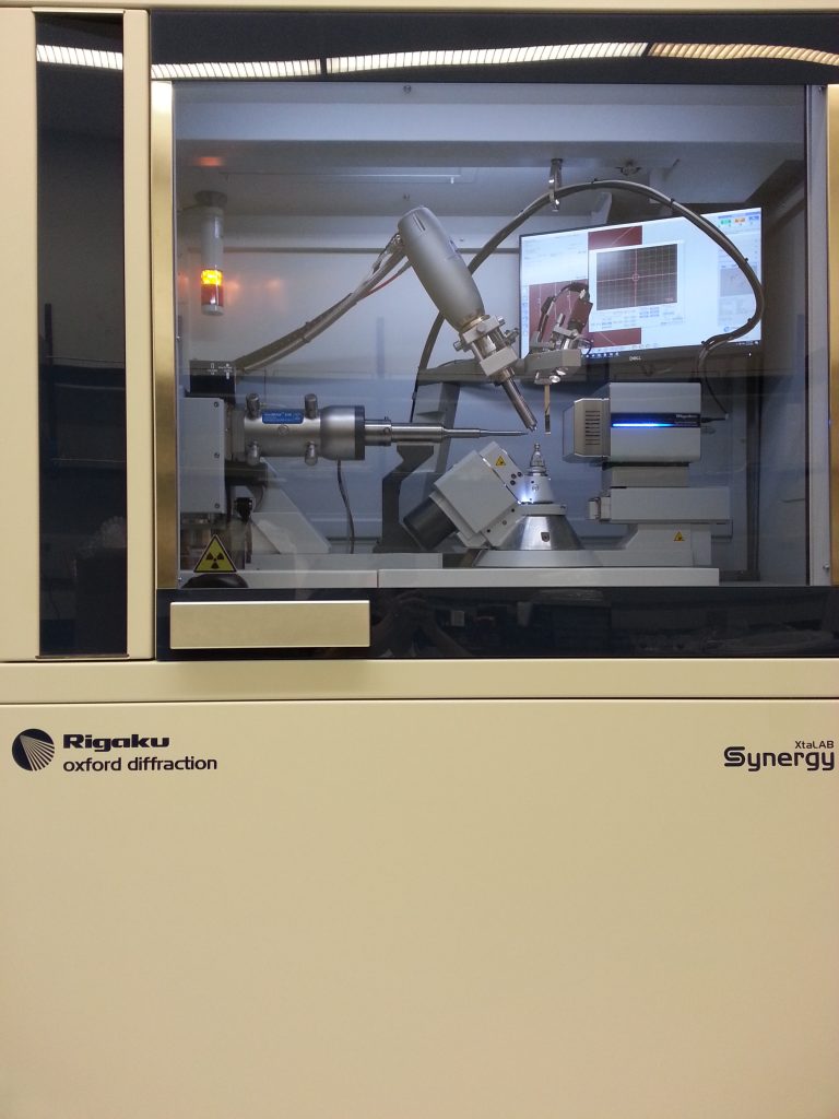
In March 2019, we retired our veteran Bruker instrument and installed the brand new Rigaku XtaLAB Synergy-R DW – Single Crystal X-Ray diffractometer (SC-XRD). The instrument is conveniently situated in Shelby Hall 3034.
The instrument features powerful X-ray beam from rotating anode with dual wavelength (Mo and Cu). The detector allows shutterless mode and has great dynamic range (high S/N ratio at both weak and strong signals). The instrument analyzes the following sample types: mineral samples, organic/organometallic samples and protein samples. Although the instrument is mainly for single crystal samples, it is also capable of analyzing powder samples with the advantage that it only requires a few mg of samples, albert with lower data quality compared to standard PXRD instrument. The typical data collection time ranges from less than one hour (for some small molecule crystals) to up to one day (for some protein samples).
The instrument is serving the needs of the chemistry department, as well as external samples. The instrument is maintained by a staff scientist with PhD degree in chemistry. Please contact the department’s main office if you have any questions.
Sample Submission
I have set up two sample boxes right outside the XRD lab door, one for sample drop-off, the other pickup. If your samples are stable, please drop off your samples (ideally with mother liquor present) together with the filled sample submission forms (one form per sample) to the drop-off box. And please do send me an email to let me know you have dropped your samples there so I can take care of them as soon as possible. If your samples are sensitive (time, air, moisture, light, temperature, etc.), please contact me in person. I will reserve a time slot with the instrument for your samples. Click here for the:
After your samples are analysed, I will drop them in the pickup box. I will send you the preliminary structure or let you know your samples are not good. It is then the time to pick up your samples. Please make sure to take back your samples in a few days. The XRD lab does not have the facilities to keep the samples for long.
Acknowledgement
Please acknowledge the new Rigaku instrument when publishing by including: “We thank NSF CHE MRI 1828078 and UA for the purchase of the single crystal X-Ray diffraction instrument.”
Please include me (Fengrui Qu) as co-author if I collected/analyzed the data for your samples.
Please inform Dr. Fengrui Qu and Dr. Elizabeth Papish of published papers acknowledging the new instrument.
Sample Fees
Chemistry UA users: Free.
Non-Chemistry UA users : $75 per structure with full analysis + report. 35$ per sample with data collection (and export) only. $30/hour for staff time on stuff that doesn’t fit those umbrellas, $20/hour for instrument dwell time that doesn’t require staff effort.
Regional PUIs as outreach : Free
Academic non-UA users (non-PUI) : $200 per structure with full analysis + report. 100$ per sample for data collection only.
Industrial samples : $400 per structure including everything (ready for publication) or, $300 per sample if you only need data collection but no data processing; 100$ for connectivity only if it is organic compound; 50$ for unit cell only. Discount are to be expected if you have more samples at once. For example, 1000$/3 samples instead of 1200$.
Possibly higher charges for macromolecule structures or any sample taking longer than one day of instrument time
Possibly a maximum charge for users, proposed at $1-2K for UA and $3-10K for industrial.
Useful Resources of Crystallography
WebCSD
This is the online searching page for CCDC database, where you can search by structure, unit cell, etc. Before you attempted to grow/submit your crystals, it is always recommended to search the database to see if the structure has been published.
checkCIF
This is where you run an online checkcif before you submit the data for publication. If your final full data set come from me, you do not need to do this. If you do your own analysis, you must go through this process to make sure your analysis is valid. The suspicious “problems” are flagged as A/B/C/G level alerts. You should try your best to eliminate A and B level alerts. Small amount of certain C level alerts are usually acceptable for most journals. But large amount of C level alerts should be examined carefully as they collectively could indicate some severe problems. Currently this tool is only good for small molecules, but not for large biomolecules.
SHELXL
This page is the homepage for SHELXL package, the heart/brain for most of today’s crystallographic programs, esp. for small molecules single crystals. It also finds its use in powder XRD and protein XRD. The package is totally free for academic users. What you only need to do is registration. Once registered, you can download the whole package to your own computer. BTW, this SHELXL package is needed before you can use any of the popular GUI programs, ,such as WinGX, Olex2, Shelxle, etc. This page also have the instructions for all the four letter cards you may use during the refinement process, such as: AFIX, DFIX, TWIN, etc.
PLATON
The Platon program is a powerful third party program which you can call from within some GUI programs (e.g., Olex2). It is very powerful and versatile. To name a few: It can find the missing symmetry (or pseudo symmetry) of your current space group. It can detect unaccounted (non)-merohedral twinning in your data. Bear in mind though it only provides reasonable suggestions. It is up to you to determine if they are valid. You can register and download the package to your local computer. Following the instructions, you can set up such that it can be called from within your favorite GUI program.
Mercury
This page has the link for you to download the powerful and free structural visualization software – Mercury. Chances are you will need this program to view your crystal structure and prepare figures for publication.
High-Resolution Space Groups
This webpage provides the space group diagrams and tables for all the 230 space groups. You can find there the symmetry operators/operations, reflection conditions, etc.
3-Dimensional Space Groups
This webpage provides another way of listing the 230 space groups, with some convenient color coding.
FAQs
You can try your best to answer all the questions you can answer and leave the rest in blank. The form is for researchers from various fields. Some of the questions may not apply to you.
Currently we are trying to minimize the instrument operators due to the delicate nature of this instrument. However, if your adviser can justify such a need to the instrument committee, we can certainly make it happen. First you need to take the crystallography course taught by Dr Jared Allred. After that, you will be trained in person by me. You can run your own samples if I think you are ready to do so.
It depends. If the instrument is available with me, I typically get started within 2 days. And I normally can give you a preliminary structure one or two day after. If the data look good, you can expect the full final data set ready for publication in about a week. If your samples are not good for whatever reason, you typically will hear from me the same day I work on your sample. Sometimes the data look promising but not good yet, for examples of disorders/twinning, etc, you will not hear from me so soon. I currently maintain the queue for the instrument time, you are welcome to check with me for your position in the queue.
It will be ideal if you can use the instrument whenever it is free. Actually that is how most users are doing currently. That way I just notify you once it is ready. The instrument get used most efficiently this way. However, if your schedule is so tight that you have to run the samples at certain times, please talk with me as soon/early as you have such needs. This way I can better coordinate the instrument time with your needs. That being said, please refrain from doing so if possible. We will try to only reserve the instrument time for courses, and urgent needs (such as sensitive samples, et al.).
I can teach you how to prepare publication figures in Mercury if you need. I can re-label the atoms if necessary, although it is recommend you provide your preferred labeling scheme during sample submission. I can combine the multiple cif files into one if you want to. I can deposit the cif files and give you the deposit numbers for you to include in the manuscript. I normally provide a detailed report accompanying the final data set. You can choose to include all or part of the report into the experimental section in your manuscript.
Note, they are all free for academic users. However, they all require registration before downloading.
For WinGX: http://www.chem.gla.ac.uk/~louis/software/wingx/
For Olex2: https://www.olexsys.org/Software
For ShelXle: https://www.shelxle.org/shelx/eingabe.php
There are tons of good tutorials on youtube if you are good at self-education. Dr Allred also covers this topic in his class. Many people in our department have experiences with at least one of the three. You may find someone in person to get started. I personally have used Olex2 and ShelXle and will be able to help you if needed
Crystal structures are obtained by Fourier transform of the diffractions, the spots you see on the frames. The final refinement was realized by full matrix least-square process. The process involves iteratively modifying all the parameters to minimize the difference between the observed diffraction intensity and the calculated intensity from the proposed model. This process normally requires over-determination, i.e., data/parameter >8. When the crystals are not diffracting strong enough. The data will be of lower quality, such as: weak spots, diffuse spots, split spots, low angle diffraction, etc. The model quality will not exceed that of the data. Overall the model will be of low quality. It could manifest in the following: The bond distances have very large esds (standard uncertainty, e.g.: 1.54(22)); The thermal factors are unusually large (the thermal ellipsoids of the atoms are very big), etc. Quite often restraints and/or constraints are implemented to ensure the model will behave. For example, “DFIX 1.54 C1 C2” will restrain the bond distance between C1-C2 to be 1.54 within stipulated tolerance. Apparently in these cases you cannot trust these values with much confidence. Rather, the structure only serves to show you the overall configration of the molecule at best. If you want to publish the structure, you will need to grow a better crystal and try again.
For small molecules, the most commonly used methods are vapor diffusion and solvent evaporation. The fundamental principle is to slowly drive the solution to over saturation, such that compound starts to seeding and then seeds start to grow. You need to control the speed of concentration changing to maintain a delicate balance between seeds formation and seeds growth. Seeds formation are usually kinetically favored. If seeds form too fast, you may end up in many tiny crystals or even precipitate. If the solution does not grow any crystals after standing for very long, you may induce crystallization by scratch the glass side wall with a glass rod, or introducing some seeding crystals with your previous less-than-ideal crystals. Remember to cut the seeding crystals to small pieces before adding. You may also vary the following conditions: preparing a series of different starting concentrations; varying solvents or solvents combinations; varying temperature; add some additives to aid crystal packing. For protein samples these rules still apply. But you need special training to be able to grow them. Please consult Dr Dunkle and Dr Thompson for details.
You may vary the solvents, temperature as described above. Growing the crystal slower and at higher temperature usually give you better crystals. Sometimes you cannot predict what alterations will bring you luck. Just try different conditions you can think of.
Crystal structure is an average representation of millions of individual molecules crystallized in the crystals. No crystals are perfect. And above zero Kelvin degree the atoms do vibrate. Small amount of thermal motion can be accounted for by the B factor (the temperature factor, also know the Debye-Waller factor). However, if the motion is very large, or the same atom actually physically occupy two different position in different unit cell, you will see two such atoms in the average image. Hence the model has disorder. Another great analogy is the 1000 soldier in a line at ease. Some are looking left, some are looking right. still some are shaking their heads. If you want to use one solder to represent all the soldiers of different heads orientation, you would have “disorder”. For the soldiers looking left or right, they are represented with static disorder. For those who are shaking their heads, it is called dynamic disorder. The latter can be mitigated by cooling to lower temperature. Yet another type of disorder is substitutional disorder, where different atoms occupy the same average position. This is more often in mineral crystals. When you are told your structures have disorder, you need to be aware its implications. If the disordered part is a small portion (by mass) of your structure and it is the non bonded solvent, then this does not negatively affect your interpretation of your major molecular structure. For that reason you should try to avoid DCM and chloroform as crystallization solvent. Also try to aovid ClO4 as counteranion if possible. If the disorders are on your major molecule of your interests, then you need to be careful when discuss the bond length and angles of the disordered part. Quite often some restraints and/or constraints are implimented during the modelling of the disorders. In such situations the discussion of the bond length and angles are meaning less. It only serves to show you the connectivity. If you do want to get the exact/accurate bond distances/angles, you need to regrow the crystal which do not show the disorders.
Twinning is a quite complicated subject. Loosely speaking, it means in the crystal packing some part(s) of the crystal have different orientations than the rest part(s), and the mutual orientation(s) can be brought to coincidence by a definitive matrix operation. The matrix operation usually are rotation around some axis, or reflection on a plane. Twining can be classified into: non-merohedral twinning, merohedral twinning, reticular obverse/reserve twinning, pseudo-merohedral twinning. Twinning will introduce difficulties at one or all the stages including: indexing, data acquisition, solving and refinement. Twinning, when taken care of properly, does not affect the structure quality (as opposed to disorders). However, if some twinning go unheeded, it will lead to spurious large Q peaks around some atoms at the best. Sometimes it causes pathological ellipsoids of atoms. Other times it will cause bond distances to be unusually long or short, which can also be caused by wrong space group assignment. The best practice is to find the best crystal you can find. And be vigilant if you see some common warning signs. Fortunately, from the perspectives of end user of the data, you do not need to worry about the twinning if the overall data quality is good. You only need to indicate the information in the experimental section, which I usually include in the final report anyway. Unlike disorders, the properly accounted twin data set is as good as non-twin single crystal data set, which you can safely discuss the structural parameters. But, if you see some unusual structural features (unusual bond distances, bond angles), do not be excited yet. Talk to the one who processed your data and have them re-examine your refinement carefully to make sure you have not missed any twinning, or assigned the space group incorrectly.
Pretty much all the solvents which have large range of solubility toward your compound will be good. However, you should try to avoid DCM and Chloroform. For one, they have heavy atoms compared to carbon atoms. Secondly, they nearly always find themselves in disorder, which will negatively affect the overall data quality of the whole crystal structure. For the sample reason, you should also try to avoid ClO4 as counteranion.
If you are close, it’s better to send samples in person. During the trip please keep the samples in an environment as close to its growth environment as possible. If you are far away or do not want to drive, you can ship the samples to me. When you pack the samples: if your samples will disintegrate upon solvent loss, you may need to keep small amount of mother liquor in the vial and seal it very tightly. Too much solvent will dissolve your crystals upon heating during the shipment. Some crystals are quite fragile and will disintegrate upon rough handling during the shipping. In that situation the only option will be to carry them over in person.
Please ship your sample to:
Dr Fengrui Qu
1048 Shelby Hall
250 Hackberry Lane
Tuscaloosa, Alabama 35487